Cardiac Investigations
The heart is a beautiful piece of bio-engineering. It has an electrical system that keeps a regular rhythm. There are valves that ensure blood flows in the right direction and the muscles contracts more than 100,000 times a day without tiring.
Unfortunately, things can go wrong and symptoms may develop. Fortunately, there are tests that can assess all aspects of the heart.
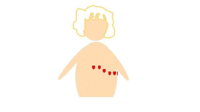
The Electrocardiogram (ECG)
Chest electrodes can be used to record the electrical signals from the heart. A low signal may be due to heart failure. A slow signal may mean heart block. The shape of the signal can identify diseases such as coronary disease. Importantly, the ECG measures the heart rate and indicates whether the rhythm is normal or abnormal as in atrial fibrillation.
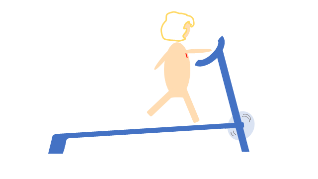
Treadmill Exercise Test
The ECG is usually recorded at rest but it can also be recorded during exercise on a treadmill. This test is often used to test the cardiovascular fitness of commercial drivers and pilots and the DVLA and CAA use it for regulatory reasons.

Event Monitor
Sometimes, cardiac symptoms are infrequent and short lasting. This makes it difficult to record an ECG at the time of symptoms but there are a number of options. There are wearable devices which can record ECGs for many days. There are also pocket devices which can be used when symptoms occur. Some devices are small enough to be implanted under the skin for several years. The latest smart watches can record ECGs.
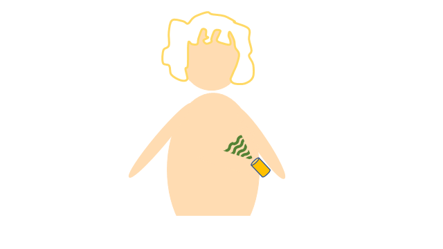
Echocardiography
Just as bats use sound to “see”, echocardiography is the use of ultrasound to investigate the structure and function of the heart. This is the same test that is used to see the baby inside a pregnant woman’s womb. It has been described as the Gold Standard test for diagnosing heart failure. Usually, the sound waves are directed towards the heart by placing a probe on the chest wall. This is known as a trans-thoracic echocardiogram.
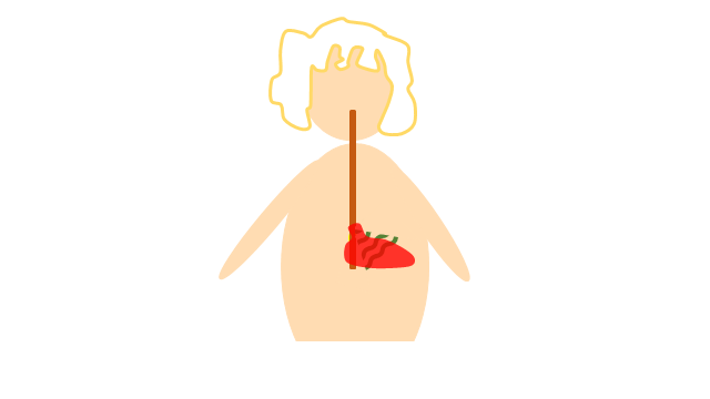
Trans-oesophageal Echo
Sometimes, the images from a transthoracic echocardiogram are not clear because ultrasound waves do not pass well through any lung tissue in the way. An ultrasound probe can be placed down the gullet which is literally just behind the heart. This is known as a trans-oesophageal echocardiogram (TOE) and the image quality is usually excellent.
Cardiac Catheterisation
In this test, narrow tubes (catheters) are inserted into an artery at the wrist or the top of a leg. The catheters are long enough to reach the heart. Dye is then injected to see the coronary arteries and to assess how well the heart pumps. Nowadays, the test is mainly done from the wrist as it is considered safer.
There are many other investigations. These include a blood test known as BNP (brain natriuretic peptide) which is used to exclude heart failure. A CT coronary angiogram is a non-invasive test to assess the coronary arteries and current guidelines recommend it as a first line test for the assessment of stable chest pain. A cardiac MR scan provides data about the structure and function of the heart as well as about the cardiac muscle. There is some overlap in the information from all these tests. Also, each test has different accuracy and some may carry risk. The decision as to which test to use will depend on factors such as patient choice and the information required.